Copyright
©The Author(s) 2016.
World J Radiol. Mar 28, 2016; 8(3): 298-307
Published online Mar 28, 2016. doi: 10.4329/wjr.v8.i3.298
Published online Mar 28, 2016. doi: 10.4329/wjr.v8.i3.298
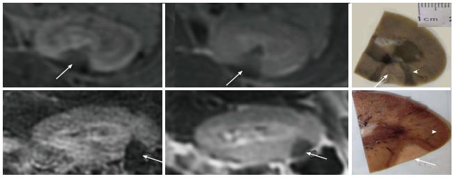
Figure 6 The wedge-shape lesions on contrast enhanced liver acquisitions with volume acquisition (left) and T1-weighted fast spin echo (center) images and gold-standard macroscopic slices (white arrows) from 2 animals (top and bottom rows).
Note the close correspondence of ablated lesions in situ and in vitro. Coagulation necrosis (white arrows) is surrounded by hemorrhagic zone (arrowheads) as shown macroscopically.
- Citation: Saeed M, Krug R, Do L, Hetts SW, Wilson MW. Renal ablation using magnetic resonance-guided high intensity focused ultrasound: Magnetic resonance imaging and histopathology assessment. World J Radiol 2016; 8(3): 298-307
- URL: https://www.wjgnet.com/1949-8470/full/v8/i3/298.htm
- DOI: https://dx.doi.org/10.4329/wjr.v8.i3.298


