Copyright
©The Author(s) 2016.
World J Radiol. Mar 28, 2016; 8(3): 298-307
Published online Mar 28, 2016. doi: 10.4329/wjr.v8.i3.298
Published online Mar 28, 2016. doi: 10.4329/wjr.v8.i3.298
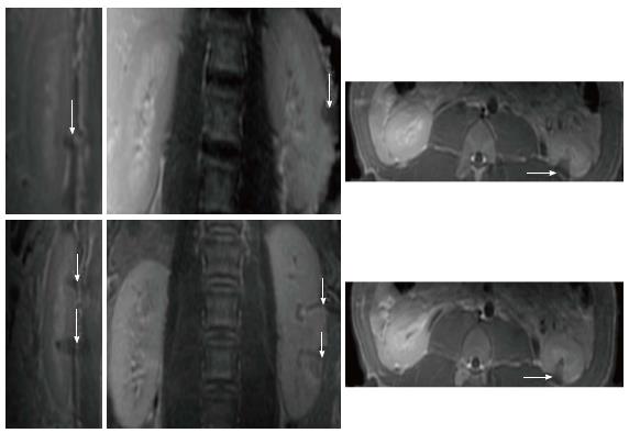
Figure 5 Renal lesions on three magnetic resonance imaging views acquired 3 h after sonication using single 4400J (top row, animal 1) and double 4400J (bottom row, animal 2).
The wedge-shape hypoenhanced lesions are seen on contrast enhanced T1-weighted fast spin echo images in sagittal (left images), coronal (center images) and axial (right images) views.
- Citation: Saeed M, Krug R, Do L, Hetts SW, Wilson MW. Renal ablation using magnetic resonance-guided high intensity focused ultrasound: Magnetic resonance imaging and histopathology assessment. World J Radiol 2016; 8(3): 298-307
- URL: https://www.wjgnet.com/1949-8470/full/v8/i3/298.htm
- DOI: https://dx.doi.org/10.4329/wjr.v8.i3.298


