Copyright
©The Author(s) 2016.
World J Radiol. Mar 28, 2016; 8(3): 288-297
Published online Mar 28, 2016. doi: 10.4329/wjr.v8.i3.288
Published online Mar 28, 2016. doi: 10.4329/wjr.v8.i3.288
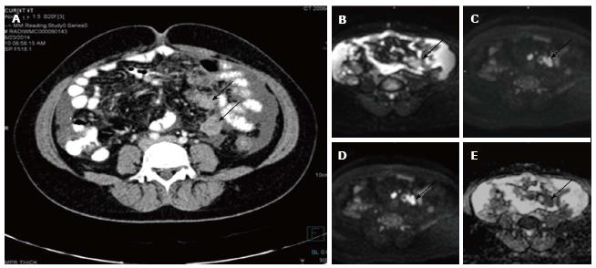
Figure 4 Serosal deposits in carcinoma ovary.
Axial contrast enhanced computed tomography image (A) of a patient with known ovarian cancer shows enhancing soft tissues (arrows) over the surface of small bowel suggestive of serosal deposits. On diffusion weighted imaging, b 0 (B), b 400 (C), b 800 (D) and ADC (E) images show significant diffusion restriction in the serosal deposits. These lesions were proven to be malignant deposits on fine needle aspiration cytology. ADC: Apparent diffusion coefficient.
- Citation: Manoharan D, Das CJ, Aggarwal A, Gupta AK. Diffusion weighted imaging in gynecological malignancies - present and future. World J Radiol 2016; 8(3): 288-297
- URL: https://www.wjgnet.com/1949-8470/full/v8/i3/288.htm
- DOI: https://dx.doi.org/10.4329/wjr.v8.i3.288


