Copyright
©The Author(s) 2016.
World J Radiol. Mar 28, 2016; 8(3): 288-297
Published online Mar 28, 2016. doi: 10.4329/wjr.v8.i3.288
Published online Mar 28, 2016. doi: 10.4329/wjr.v8.i3.288
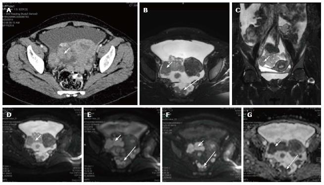
Figure 3 Carcinoma ovary.
Axial contrast enhanced computed tomography image (A), T2 weighted axial (B) and coronal (C) images of a patient with known ovarian cancer shows right adnexal mass (arrows) multiple deposits in peritoneum of the pelvis (long arrows); On DWI, b 0 (D), b 400 (E), b 800 (F) and ADC (G) images show significant diffusion restriction in the primary (arrow) as well as peritoneal deposits (long arrow). These lesions were proven to be malignant deposits on fine needle aspiration cytology. ADC: Apparent diffusion coefficient; DWI: Diffusion weighted imaging.
- Citation: Manoharan D, Das CJ, Aggarwal A, Gupta AK. Diffusion weighted imaging in gynecological malignancies - present and future. World J Radiol 2016; 8(3): 288-297
- URL: https://www.wjgnet.com/1949-8470/full/v8/i3/288.htm
- DOI: https://dx.doi.org/10.4329/wjr.v8.i3.288


