Copyright
©The Author(s) 2016.
World J Radiol. Mar 28, 2016; 8(3): 288-297
Published online Mar 28, 2016. doi: 10.4329/wjr.v8.i3.288
Published online Mar 28, 2016. doi: 10.4329/wjr.v8.i3.288
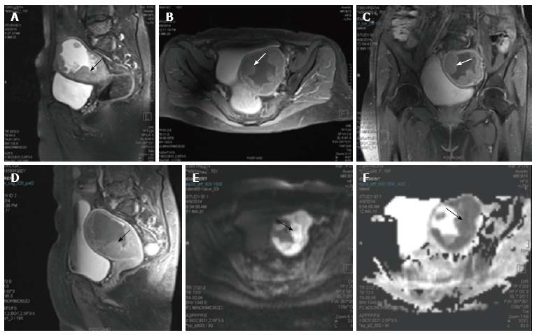
Figure 2 Endometrial carcinoma.
Sagittal T2 weighted magnetic resonance image of a female who presented with menorrhagia. It shows irregular nodular thickening of the endometrial (arrow in A). The lesion on post contrast axial (B), coronal (C) and sagittal (D) images shows enhancement. On diffusion weighted imaging (E) and corresponding apparent diffusion coefficient (F) images, the lesion showed significant restriction of diffusion. The tumour is limited to the outer myometrium and no parametrial invasion noted suggesting a T1c disease. This lesion on biopsy turned out to be carcinoma endometrium.
- Citation: Manoharan D, Das CJ, Aggarwal A, Gupta AK. Diffusion weighted imaging in gynecological malignancies - present and future. World J Radiol 2016; 8(3): 288-297
- URL: https://www.wjgnet.com/1949-8470/full/v8/i3/288.htm
- DOI: https://dx.doi.org/10.4329/wjr.v8.i3.288


