Copyright
©The Author(s) 2016.
World J Radiol. Mar 28, 2016; 8(3): 275-280
Published online Mar 28, 2016. doi: 10.4329/wjr.v8.i3.275
Published online Mar 28, 2016. doi: 10.4329/wjr.v8.i3.275
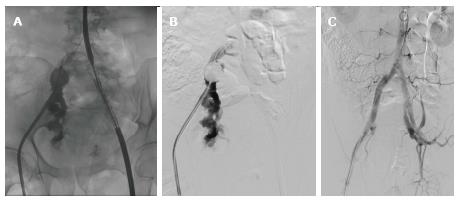
Figure 4 Rupture of the right external iliac artery during manipulation.
A, B: Partially deployed IBD on the left side. An occlusive balloon placed in the right common iliac artery. Angiography revealed the rupture site at the proximal part of the external iliac artery; C: Control angiography following placement of the two fluency stent grafts on the right side, and completion of the procedure with the placement of an IBD device on the left side. IBD: Iliac branch device.
- Citation: Duvnjak S. Endovascular treatment of aortoiliac aneurysms: From intentional occlusion of the internal iliac artery to branch iliac stent graft. World J Radiol 2016; 8(3): 275-280
- URL: https://www.wjgnet.com/1949-8470/full/v8/i3/275.htm
- DOI: https://dx.doi.org/10.4329/wjr.v8.i3.275


