Copyright
©The Author(s) 2016.
World J Radiol. Mar 28, 2016; 8(3): 268-274
Published online Mar 28, 2016. doi: 10.4329/wjr.v8.i3.268
Published online Mar 28, 2016. doi: 10.4329/wjr.v8.i3.268
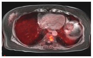
Figure 3 A 63-year-old female with renal failure and abnormal serum protein electrophoresis.
Positron emission tomography-magnetic resonance imaging revealed focal abnormal heterogeneously T1 hypointense and T2 hyperintense lesion with corresponding increased FDG uptake. Lesion biopsy was positive for multiple myeloma. FDG: 18F-fluorodeoxyglucose.
- Citation: Chaudhry AA, Gul M, Gould E, Teng M, Baker K, Matthews R. Utility of positron emission tomography-magnetic resonance imaging in musculoskeletal imaging. World J Radiol 2016; 8(3): 268-274
- URL: https://www.wjgnet.com/1949-8470/full/v8/i3/268.htm
- DOI: https://dx.doi.org/10.4329/wjr.v8.i3.268


