Copyright
©The Author(s) 2016.
World J Radiol. Mar 28, 2016; 8(3): 240-254
Published online Mar 28, 2016. doi: 10.4329/wjr.v8.i3.240
Published online Mar 28, 2016. doi: 10.4329/wjr.v8.i3.240
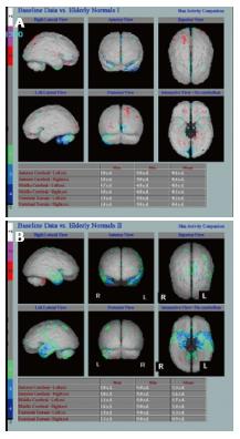
Figure 4 Underlying neuronal dysfunction and neurodegeneration.
A: Hexamethylpropylene amine oxime (HMPAO) single photon emission computed tomography (SPECT) in normal control subject demonstrating normal almost symmetrical perfusion pattern; B: HMPAO SPECT in Alzheimer’s disease parametric images demonstrate bilateral reduction in perfusion in the temporal lobes especially in the medial temporal regions up to 2 (green) and 3 (blue) standard deviation (Images kindly prepared by Ms Lesley Lovell, Senior technician).
- Citation: Narayanan L, Murray AD. What can imaging tell us about cognitive impairment and dementia? World J Radiol 2016; 8(3): 240-254
- URL: https://www.wjgnet.com/1949-8470/full/v8/i3/240.htm
- DOI: https://dx.doi.org/10.4329/wjr.v8.i3.240


