Copyright
©The Author(s) 2016.
World J Radiol. Mar 28, 2016; 8(3): 240-254
Published online Mar 28, 2016. doi: 10.4329/wjr.v8.i3.240
Published online Mar 28, 2016. doi: 10.4329/wjr.v8.i3.240
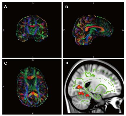
Figure 2 Diffusion tensor imaging.
A-C: Diffusion tensor imaging (DTI) data set superimposed on structural image of the brain in 3 orthogonal planes demonstrating colour coded white matter tracts. Blue colour correlate to the tracts in the cranio-caudal direction, red in the transverse direction and green in the antero-posterior direction. (Images kindly prepared by Dr. Gordon D Waiter); D: DTI data of white matter tracts (green) superimposed on T1 image demonstrating statistically significant difference in fractional anisotropy in the fornix (orange areas) compared to the rest of the brain in a subgroup of patients. (Images kindly prepared by Dr. Gordon D Waiter).
- Citation: Narayanan L, Murray AD. What can imaging tell us about cognitive impairment and dementia? World J Radiol 2016; 8(3): 240-254
- URL: https://www.wjgnet.com/1949-8470/full/v8/i3/240.htm
- DOI: https://dx.doi.org/10.4329/wjr.v8.i3.240


