Copyright
©The Author(s) 2016.
World J Radiol. Mar 28, 2016; 8(3): 226-239
Published online Mar 28, 2016. doi: 10.4329/wjr.v8.i3.226
Published online Mar 28, 2016. doi: 10.4329/wjr.v8.i3.226
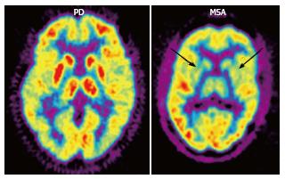
Figure 4 2-[18F]-fluoro-2-deoxy-D-glucose positron emission tomography (FDG PET) in Parkinson’s disease (PD) and multiple system atrophy (MSA).
FDG PET shows hypometabolism in bilateral parieto-occipital and prefrontal cortices, as well as hypermetabolism in the basal ganglia and thalamus in PD, as opposed to bilateral striatal and thalamic hypometabolism in MSA (arrows). (Originally published in the JNM. Brooks DJ. J Nucl Med 2010; 51: 596-609. © by the Society of Nuclear Medicine and Molecular Imaging, Inc.)
- Citation: Lizarraga KJ, Gorgulho A, Chen W, De Salles AA. Molecular imaging of movement disorders. World J Radiol 2016; 8(3): 226-239
- URL: https://www.wjgnet.com/1949-8470/full/v8/i3/226.htm
- DOI: https://dx.doi.org/10.4329/wjr.v8.i3.226


