Copyright
©The Author(s) 2016.
World J Radiol. Mar 28, 2016; 8(3): 226-239
Published online Mar 28, 2016. doi: 10.4329/wjr.v8.i3.226
Published online Mar 28, 2016. doi: 10.4329/wjr.v8.i3.226
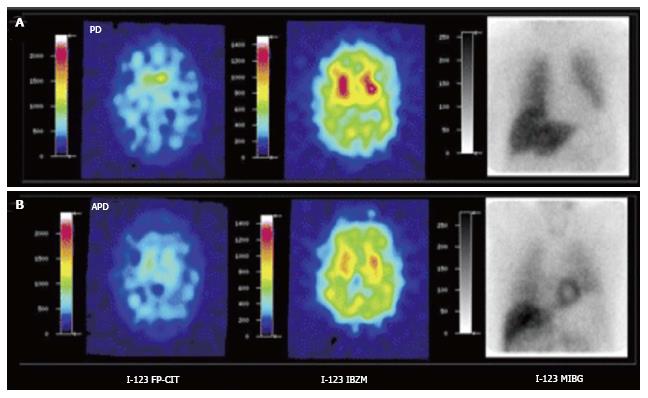
Figure 3 [123I]FPCIT, [123I]IBZM and cardiac SPECT imaging [[123I]MIBG] of Parkinson’s disease (A) and atypical parkinsonism (B).
Striatal [123I]FPCIT is decreased in both PD and APD. Striatal D2 receptor binding is normal in PD but decreased in APD. Myocardial sympathetic SPECT signal is decreased in PD but normal in APD. PD: Parkinson’s disease; APD: Atypical parkinsonism; SPECT: Single photon emission computerized tomography. (Originally published in the JNM. Südmeyer M, Antke C, Zizek T, et al. J Nucl Med 2011; 52: 733-740. © by the Society of Nuclear Medicine and Molecular Imaging, Inc.)
- Citation: Lizarraga KJ, Gorgulho A, Chen W, De Salles AA. Molecular imaging of movement disorders. World J Radiol 2016; 8(3): 226-239
- URL: https://www.wjgnet.com/1949-8470/full/v8/i3/226.htm
- DOI: https://dx.doi.org/10.4329/wjr.v8.i3.226


