Copyright
©The Author(s) 2016.
World J Radiol. Mar 28, 2016; 8(3): 226-239
Published online Mar 28, 2016. doi: 10.4329/wjr.v8.i3.226
Published online Mar 28, 2016. doi: 10.4329/wjr.v8.i3.226
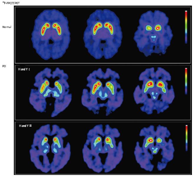
Figure 2 Nigrostriatal dopaminergic system degeneration in Parkinson’s disease.
PET images show striatal uptake of [18F]FPCIT, a ligand with high affinity for pre-synaptic dopamine transporters, for a normal volunteer (top row), and 2 patients with Parkinson’s disease (PD) [middle row: Hoehn and Yahr (H and Y) stage I; bottom row: H and Y stage III]. Striatal [18F]FPCIT uptake is mainly reduced in the putamen contralateral to the most symptomatic side in early PD (middle row), progressing to bilateral reduction in a caudal-to-rostral pattern (bottom row). (Originally published in the JNM. Katzumata K, Dhawan V, Chaly T, et al. Dopamine transporter imaging with fluorine-18-FPCIT and PET. J Nucl Med 1998; 39: 1521-1530. © by the Society of Nuclear Medicine and Molecular Imaging, Inc.)
- Citation: Lizarraga KJ, Gorgulho A, Chen W, De Salles AA. Molecular imaging of movement disorders. World J Radiol 2016; 8(3): 226-239
- URL: https://www.wjgnet.com/1949-8470/full/v8/i3/226.htm
- DOI: https://dx.doi.org/10.4329/wjr.v8.i3.226


