Copyright
©The Author(s) 2016.
World J Radiol. Feb 28, 2016; 8(2): 200-209
Published online Feb 28, 2016. doi: 10.4329/wjr.v8.i2.200
Published online Feb 28, 2016. doi: 10.4329/wjr.v8.i2.200
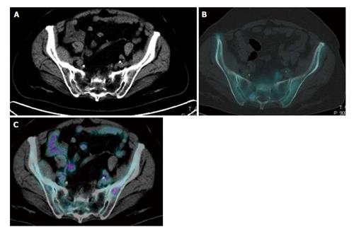
Figure 2 Evidence of the complementary features of 2-deoxy-2-(18F)fluoro-D-glucose and 18F-sodium positron emission tomography/computed tomography in the pelvis of the same breast cancer patient.
An 18F-NaF avid sclerotic lesion was detected in the right sacrum in absence of significant 18F-FDG uptake. By contrast high uptake of 18F-FDG was present in a small lytic lesion in the left iliac bone. Due to the absence of a significant local bone reaction, this small lesion did not show any uptake of 18F-NaF. Both lesions corresponded to metastatic sites of disease and disappeared after chemotherapy. 18F-FDG: 2-deoxy-2-(18F)fluoro-D-glucose; 18F-NaF: 18F-sodium; PET/CT: Positron emission tomography/computed tomography.
- Citation: Capitanio S, Bongioanni F, Piccardo A, Campus C, Gonella R, Tixi L, Naseri M, Pennone M, Altrinetti V, Buschiazzo A, Bossert I, Fiz F, Bruno A, DeCensi A, Sambuceti G, Morbelli S. Comparisons between glucose analogue 2-deoxy-2-(18F)fluoro-D-glucose and 18F-sodium fluoride positron emission tomography/computed tomography in breast cancer patients with bone lesions. World J Radiol 2016; 8(2): 200-209
- URL: https://www.wjgnet.com/1949-8470/full/v8/i2/200.htm
- DOI: https://dx.doi.org/10.4329/wjr.v8.i2.200


