Copyright
©The Author(s) 2016.
World J Radiol. Feb 28, 2016; 8(2): 174-182
Published online Feb 28, 2016. doi: 10.4329/wjr.v8.i2.174
Published online Feb 28, 2016. doi: 10.4329/wjr.v8.i2.174
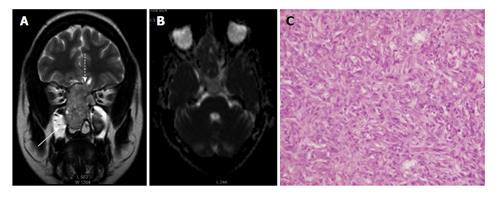
Figure 6 Sinonasal hemangiopericytoma in a 45-year-old lady.
A: Coronal T2W image shows mildly expansile sinonasal mass involving, extending to bilateral ethmoid sinuses. Small intracranial extension noted in anterior cranial fossa (dashed arrow); Retained secretions in right maxillary sinus (solid arrow); B: ADC map shows restricted diffusion in the mass; C: Photomicrograph shows spindle cells with mild pleomorphism (H-E; original magnification: × 100). ADC: Apparent diffusion coefficient.
- Citation: Das A, Bhalla AS, Sharma R, Kumar A, Sharma M, Gamanagatti S, Thakar A, Sharma S. Benign neck masses showing restricted diffusion: Is there a histological basis for discordant behavior? World J Radiol 2016; 8(2): 174-182
- URL: https://www.wjgnet.com/1949-8470/full/v8/i2/174.htm
- DOI: https://dx.doi.org/10.4329/wjr.v8.i2.174


