Copyright
©The Author(s) 2016.
World J Radiol. Feb 28, 2016; 8(2): 174-182
Published online Feb 28, 2016. doi: 10.4329/wjr.v8.i2.174
Published online Feb 28, 2016. doi: 10.4329/wjr.v8.i2.174
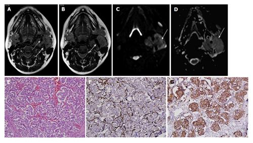
Figure 3 Carotid body tumour in a 30-year-old lady.
A and B: Axial T2W images show heterogeneously hyperintense mass in left carotid space splaying the bifurcation of left common carotid artery (arrow in B) with encasement of both ECA and ICA (dashed arrow and solid arrow in A, respectively); C and D: DWI at b500 s/mm² (C) and ADC map (D) show restricted diffusion in the mass; E: Photomicrographs show tumor cells arranged in Zellballen pattern separated by thin fibrovascular septae (H-E; original magnification: × 200); F and G: S-100 immunostain demonstrating prominence of sustentacular cells at the periphery of the tumor cell nests (F, original magnification, × 200) and tumor cells are immunopositive for synaptophysin (G, original magnification: × 200). DWI: Diffusion weighted imaging; ADC: Apparent diffusion coefficient; ECA: External carotid artery; ICA: Internal carotid artery.
- Citation: Das A, Bhalla AS, Sharma R, Kumar A, Sharma M, Gamanagatti S, Thakar A, Sharma S. Benign neck masses showing restricted diffusion: Is there a histological basis for discordant behavior? World J Radiol 2016; 8(2): 174-182
- URL: https://www.wjgnet.com/1949-8470/full/v8/i2/174.htm
- DOI: https://dx.doi.org/10.4329/wjr.v8.i2.174


