Copyright
©The Author(s) 2016.
World J Radiol. Feb 28, 2016; 8(2): 159-173
Published online Feb 28, 2016. doi: 10.4329/wjr.v8.i2.159
Published online Feb 28, 2016. doi: 10.4329/wjr.v8.i2.159
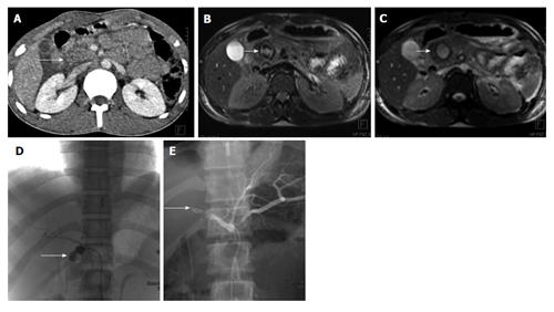
Figure 15 Post-traumatic common hepatic artery pseudoaneurysm 22 years old man with blunt trauma abdomen.
CECT axial image shows injury to head of pancreas (arrow A). MRI done 3 d later showed well defined lesion in head of pancreas with heterogeneous signal intensity on T1 weighted image (arrow B) and hyperintense on TRUFISP image (arrow C). A possibility of pseudoaneurysm was given. Angiogram showed pseudoaneurysm arising from proximal common hepatic artery (arrow D) which was embolised with Nester coils (arrow E). CECT: Contrast enhanced computed tomography; MRI: Magnetic resonance imaging; TRUFISP: True fast imaging with steady-state free precession.
- Citation: Kumar A, Panda A, Gamanagatti S. Blunt pancreatic trauma: A persistent diagnostic conundrum? World J Radiol 2016; 8(2): 159-173
- URL: https://www.wjgnet.com/1949-8470/full/v8/i2/159.htm
- DOI: https://dx.doi.org/10.4329/wjr.v8.i2.159


