Copyright
©The Author(s) 2016.
World J Radiol. Feb 28, 2016; 8(2): 159-173
Published online Feb 28, 2016. doi: 10.4329/wjr.v8.i2.159
Published online Feb 28, 2016. doi: 10.4329/wjr.v8.i2.159
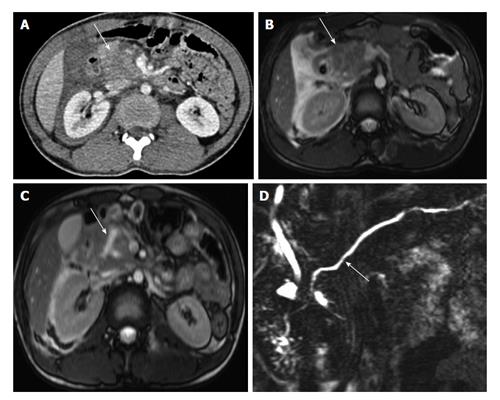
Figure 11 A 22-year-old man with history of fall of heavy object over abdomen.
CECT axial image (A) shows bulky heterogeneously attenuating head of pancreas (arrow). MRI done 28 h after CT (B) shows similar findings with bulky head and altered signal intensity with adjacent fluid (arrow B). No definite laceration seen. Follow-up MRI on day 6 (C) shows a Y shaped laceration in inferior part of head and uncinate process. However the main pancreatic duct was normal (arrow D). Since the MPD was not involved, conservative management was continued. CECT: Contrast enhanced computed tomography; MRI: Magnetic resonance imaging; CT: Computed tomography.
- Citation: Kumar A, Panda A, Gamanagatti S. Blunt pancreatic trauma: A persistent diagnostic conundrum? World J Radiol 2016; 8(2): 159-173
- URL: https://www.wjgnet.com/1949-8470/full/v8/i2/159.htm
- DOI: https://dx.doi.org/10.4329/wjr.v8.i2.159


