Copyright
©The Author(s) 2016.
World J Radiol. Feb 28, 2016; 8(2): 159-173
Published online Feb 28, 2016. doi: 10.4329/wjr.v8.i2.159
Published online Feb 28, 2016. doi: 10.4329/wjr.v8.i2.159
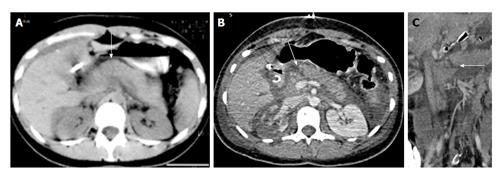
Figure 10 A 23-year-old woman with history of road traffic accident.
Day 1 CECT axial image (A) shows ill-defined contusion in pancreatic neck (arrow). No obvious laceration was seen. Day 3 CECT axial (B) and coronal oblique (C) images show a full-thickness laceration in neck of pancreas s/o grade III injury with ductal involvement. Patient was operated and distal pancreatectomy was done. Thus there was evolution of injury from contusion to laceration. CECT: Contrast enhanced computed tomography.
- Citation: Kumar A, Panda A, Gamanagatti S. Blunt pancreatic trauma: A persistent diagnostic conundrum? World J Radiol 2016; 8(2): 159-173
- URL: https://www.wjgnet.com/1949-8470/full/v8/i2/159.htm
- DOI: https://dx.doi.org/10.4329/wjr.v8.i2.159


