Copyright
©The Author(s) 2016.
World J Radiol. Feb 28, 2016; 8(2): 159-173
Published online Feb 28, 2016. doi: 10.4329/wjr.v8.i2.159
Published online Feb 28, 2016. doi: 10.4329/wjr.v8.i2.159
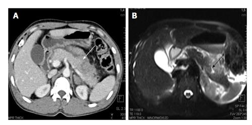
Figure 7 A 25-year-old man with history of blunt trauma abdomen.
CECT axial image (A) shows injury of distal pancreas (white arrow). MRI T2 HASTE axial image show T2 weighted hyperintensity in pancreatic body suggestive of contusion/edema with a small laceration (black arrow). CECT: Contrast enhanced computed tomography; MRI: Magnetic resonance imaging.
- Citation: Kumar A, Panda A, Gamanagatti S. Blunt pancreatic trauma: A persistent diagnostic conundrum? World J Radiol 2016; 8(2): 159-173
- URL: https://www.wjgnet.com/1949-8470/full/v8/i2/159.htm
- DOI: https://dx.doi.org/10.4329/wjr.v8.i2.159


