Copyright
©The Author(s) 2016.
World J Radiol. Feb 28, 2016; 8(2): 159-173
Published online Feb 28, 2016. doi: 10.4329/wjr.v8.i2.159
Published online Feb 28, 2016. doi: 10.4329/wjr.v8.i2.159
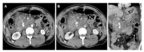
Figure 6 A 40-year-old man with history of fall of heavy object over abdomen.
CECT axial images, initial scan (A) and delayed scan (B) show complete disruption of head of pancreas with a large retroperitoneal hematoma replacing the head region. There is active extravasation of contrast (arrows). On coronal oblique image (C), the disrupted head of pancreas (arrow) with active contrast extravasation can be seen. Surgically, a crush injury (grade V) was confirmed and patient underwent Whipple’s procedure. The patient eventually died due to sepsis and multiorgan failure. CECT: Contrast enhanced computed tomography.
- Citation: Kumar A, Panda A, Gamanagatti S. Blunt pancreatic trauma: A persistent diagnostic conundrum? World J Radiol 2016; 8(2): 159-173
- URL: https://www.wjgnet.com/1949-8470/full/v8/i2/159.htm
- DOI: https://dx.doi.org/10.4329/wjr.v8.i2.159


