Copyright
©The Author(s) 2016.
World J Radiol. Feb 28, 2016; 8(2): 159-173
Published online Feb 28, 2016. doi: 10.4329/wjr.v8.i2.159
Published online Feb 28, 2016. doi: 10.4329/wjr.v8.i2.159
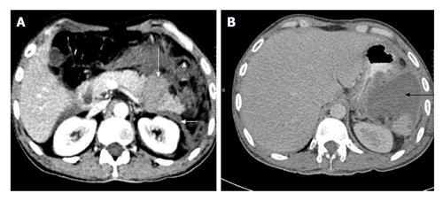
Figure 5 A 35-year-old man with road traffic accident.
CECT axial image at time of trauma (A) shows distal transection with fragments that separated by hyperattenuating fluid suggestive of hematoma (long white arrow). The left anterior renal fascia is also thickened (arrowhead). Patient underwent distal pancreatectomy and 4 wk follow up CECT axial image (B) show a post-operative collection in lesser sac (black arrow). CECT: Contrast enhanced computed tomography.
- Citation: Kumar A, Panda A, Gamanagatti S. Blunt pancreatic trauma: A persistent diagnostic conundrum? World J Radiol 2016; 8(2): 159-173
- URL: https://www.wjgnet.com/1949-8470/full/v8/i2/159.htm
- DOI: https://dx.doi.org/10.4329/wjr.v8.i2.159


