Copyright
©The Author(s) 2016.
World J Radiol. Feb 28, 2016; 8(2): 132-141
Published online Feb 28, 2016. doi: 10.4329/wjr.v8.i2.132
Published online Feb 28, 2016. doi: 10.4329/wjr.v8.i2.132
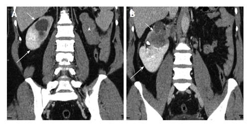
Figure 11 A 42-year-old man with crossed fused ectopia on the right side.
Coronal contrast enhanced computed tomography (A and B) shows empty left renal fossa (arrowhead) with crossed fused ectopic left kidney (long arrow) fused with the right kidney (short arrow) which shows infiltrating transitional cell carcinoma.
- Citation: Ramanathan S, Kumar D, Khanna M, Al Heidous M, Sheikh A, Virmani V, Palaniappan Y. Multi-modality imaging review of congenital abnormalities of kidney and upper urinary tract. World J Radiol 2016; 8(2): 132-141
- URL: https://www.wjgnet.com/1949-8470/full/v8/i2/132.htm
- DOI: https://dx.doi.org/10.4329/wjr.v8.i2.132


