Copyright
©The Author(s) 2016.
World J Radiol. Feb 28, 2016; 8(2): 132-141
Published online Feb 28, 2016. doi: 10.4329/wjr.v8.i2.132
Published online Feb 28, 2016. doi: 10.4329/wjr.v8.i2.132
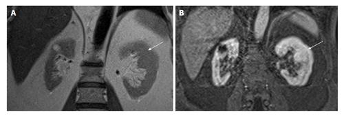
Figure 5 A 60-year-old man with suspicious mass in the left kidney on ultrasonogram.
Coronal T2W (A) and contrast enhanced T1W magnetic resonance imaging (B) shows hypertrophied column of Bertin in interpolar region of LK (arrows). LK: Left kidney.
- Citation: Ramanathan S, Kumar D, Khanna M, Al Heidous M, Sheikh A, Virmani V, Palaniappan Y. Multi-modality imaging review of congenital abnormalities of kidney and upper urinary tract. World J Radiol 2016; 8(2): 132-141
- URL: https://www.wjgnet.com/1949-8470/full/v8/i2/132.htm
- DOI: https://dx.doi.org/10.4329/wjr.v8.i2.132


