Copyright
©The Author(s) 2016.
World J Radiol. Feb 28, 2016; 8(2): 132-141
Published online Feb 28, 2016. doi: 10.4329/wjr.v8.i2.132
Published online Feb 28, 2016. doi: 10.4329/wjr.v8.i2.132
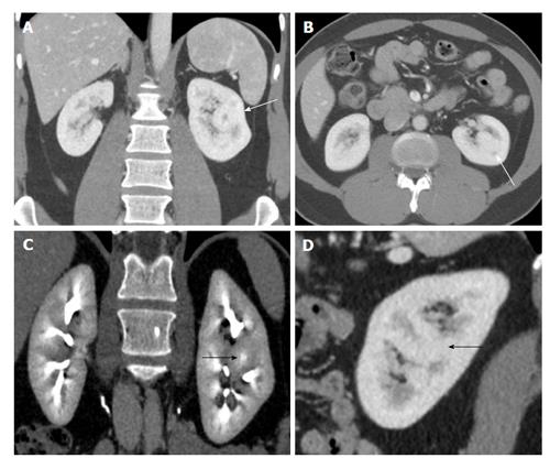
Figure 4 A 50-year-old woman with suspicious solid mass in left kidney on ultrasonogram.
Coronal (A and C), axial (B) and sagittal (D) computed tomography showing hypertrophied column of Bertin in interpolar region of LK (arrows) showing density and enhancement pattern similar to adjacent normal renal cortex. LK: Left kidney.
- Citation: Ramanathan S, Kumar D, Khanna M, Al Heidous M, Sheikh A, Virmani V, Palaniappan Y. Multi-modality imaging review of congenital abnormalities of kidney and upper urinary tract. World J Radiol 2016; 8(2): 132-141
- URL: https://www.wjgnet.com/1949-8470/full/v8/i2/132.htm
- DOI: https://dx.doi.org/10.4329/wjr.v8.i2.132


