Copyright
©The Author(s) 2016.
World J Radiol. Feb 28, 2016; 8(2): 132-141
Published online Feb 28, 2016. doi: 10.4329/wjr.v8.i2.132
Published online Feb 28, 2016. doi: 10.4329/wjr.v8.i2.132
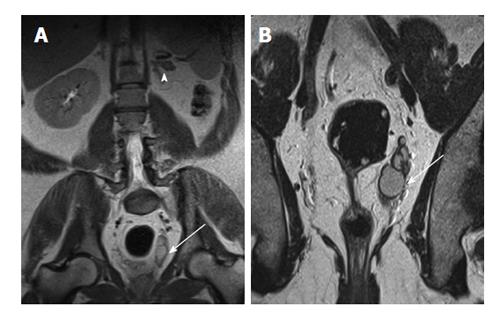
Figure 2 A 40-year-old man with left renal agenesis.
Coronal T2W magnetic resonance imaging abdomen (A) and pelvis (B) shows absent left kidney (arrowhead) and left seminal vesicle cyst (arrow).
- Citation: Ramanathan S, Kumar D, Khanna M, Al Heidous M, Sheikh A, Virmani V, Palaniappan Y. Multi-modality imaging review of congenital abnormalities of kidney and upper urinary tract. World J Radiol 2016; 8(2): 132-141
- URL: https://www.wjgnet.com/1949-8470/full/v8/i2/132.htm
- DOI: https://dx.doi.org/10.4329/wjr.v8.i2.132


