Copyright
©The Author(s) 2016.
World J Radiol. Feb 28, 2016; 8(2): 124-131
Published online Feb 28, 2016. doi: 10.4329/wjr.v8.i2.124
Published online Feb 28, 2016. doi: 10.4329/wjr.v8.i2.124
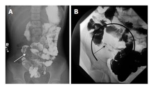
Figure 1 Small bowel follow-through examination in two patients with Crohn’s disease.
A and B demonstrate mucosal irregularity and luminal narrowing of the terminal ileum (arrows).
- Citation: Haas K, Rubesova E, Bass D. Role of imaging in the evaluation of inflammatory bowel disease: How much is too much? World J Radiol 2016; 8(2): 124-131
- URL: https://www.wjgnet.com/1949-8470/full/v8/i2/124.htm
- DOI: https://dx.doi.org/10.4329/wjr.v8.i2.124


