Copyright
©The Author(s) 2016.
World J Radiol. Feb 28, 2016; 8(2): 117-123
Published online Feb 28, 2016. doi: 10.4329/wjr.v8.i2.117
Published online Feb 28, 2016. doi: 10.4329/wjr.v8.i2.117
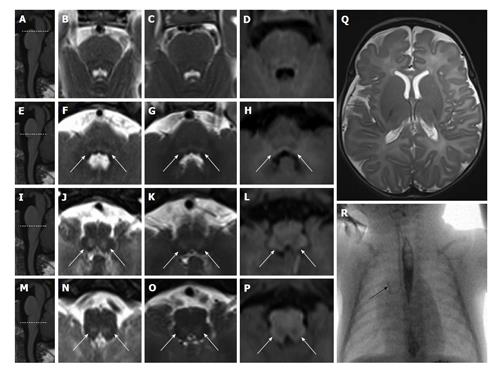
Figure 2 Brainstem tegmental lesions and oral motor dysfunction.
An infant with perinatal asphyxia due to knotting of umbilical cord around the neck is shown. Apgar score at five minutes after birth. Eighteen days after birth (panels B, F, J and N), MR images show faint T2 hyperintense (white arrows in F, J, and N) bilateral and symmetric tegmental lesions of the caudal pons and medulla oblongata. Forty days after birth (panels C, D, G, H, K, L, O and P), MR images confirm T2 hyperintense (white arrows in G, K, and O) and T1 hypointense (white arrows in H, L, and P) bilateral and symmetric tegmental lesions of the caudal pons and medulla oblongata. No signal alterations are detected at the level of the cranial pons (B-D) and at supratentorial level (Q). At 1 mo, an upper GI tract X-ray showed iodinated contrast (Iopamidol, IOPAMIRO 300) inhalation (black arrow in panel R points to the right bronchus). Gastrostomy was performed. At the age of 2 years, psychomotor delay and dysphagia are present. Sagittal views of the brainstem (panels A, E, I and M) are used for reference of axial images. MR: Magnetic resonance.
- Citation: Quattrocchi CC, Fariello G, Longo D. Brainstem tegmental lesions in neonates with hypoxic-ischemic encephalopathy: Magnetic resonance diagnosis and clinical outcome. World J Radiol 2016; 8(2): 117-123
- URL: https://www.wjgnet.com/1949-8470/full/v8/i2/117.htm
- DOI: https://dx.doi.org/10.4329/wjr.v8.i2.117


