Copyright
©The Author(s) 2016.
World J Radiol. Dec 28, 2016; 8(12): 928-932
Published online Dec 28, 2016. doi: 10.4329/wjr.v8.i12.928
Published online Dec 28, 2016. doi: 10.4329/wjr.v8.i12.928
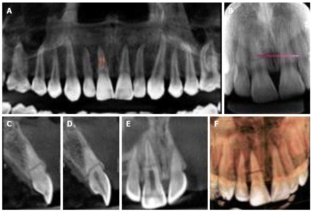
Figure 3 Cone beam tomography.
A: Panoramic coronal reconstruction of the maxilla; B: Periapical Radiograph, showing horizontal root fracture in the middle third of tooth 11; C and D: Sagital reconstructions of tooth 11, from mesial to distal sequence; E: Coronal reconstructions of the upper incisors, from buccal to lingual direction; F: 3D reconstructions (buccal and lingual views).
- Citation: Silva L, Álvares P, Arruda JA, Silva LV, Rodrigues C, Sobral APV, Silveira M. Horizontally root fractured teeth with pulpal vitality - two case reports. World J Radiol 2016; 8(12): 928-932
- URL: https://www.wjgnet.com/1949-8470/full/v8/i12/928.htm
- DOI: https://dx.doi.org/10.4329/wjr.v8.i12.928


