Copyright
©The Author(s) 2016.
World J Radiol. Dec 28, 2016; 8(12): 928-932
Published online Dec 28, 2016. doi: 10.4329/wjr.v8.i12.928
Published online Dec 28, 2016. doi: 10.4329/wjr.v8.i12.928
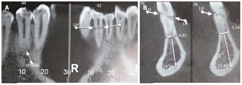
Figure 2 Cone beam tomography images.
A: Cone beam computed tomography (CT) shows apex remodeling and root canal apical third obliteration of teeth 44 and 43, as well as fracture line in tooth 42; B: Cone beam CT shows oblique fracture line along the middle third of tooth 42. LH: Horizontal line.
- Citation: Silva L, Álvares P, Arruda JA, Silva LV, Rodrigues C, Sobral APV, Silveira M. Horizontally root fractured teeth with pulpal vitality - two case reports. World J Radiol 2016; 8(12): 928-932
- URL: https://www.wjgnet.com/1949-8470/full/v8/i12/928.htm
- DOI: https://dx.doi.org/10.4329/wjr.v8.i12.928


