Copyright
©The Author(s) 2016.
World J Radiol. Dec 28, 2016; 8(12): 928-932
Published online Dec 28, 2016. doi: 10.4329/wjr.v8.i12.928
Published online Dec 28, 2016. doi: 10.4329/wjr.v8.i12.928
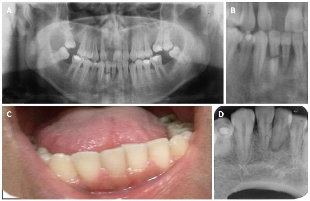
Figure 1 Radiographic and clinical as immobilize the tooth with a semi-rigid splint for 4 wk up to 4 mo.
A: Panoramic radiograph taken at the moment of the accident; B: Close at the fractured tooth at the time of the accident; C: Intraoral view. Tooth 41 was slightly grayish while tooth 42 had normal color and no mobility was present; D: Periapical radiograph, tooth 41 with a radiolucent lesion compatible with periapical lesion; tooth 42 with a thin radiolucent line at the fractured line.
- Citation: Silva L, Álvares P, Arruda JA, Silva LV, Rodrigues C, Sobral APV, Silveira M. Horizontally root fractured teeth with pulpal vitality - two case reports. World J Radiol 2016; 8(12): 928-932
- URL: https://www.wjgnet.com/1949-8470/full/v8/i12/928.htm
- DOI: https://dx.doi.org/10.4329/wjr.v8.i12.928


