Copyright
©The Author(s) 2016.
World J Radiol. Dec 28, 2016; 8(12): 922-927
Published online Dec 28, 2016. doi: 10.4329/wjr.v8.i12.922
Published online Dec 28, 2016. doi: 10.4329/wjr.v8.i12.922
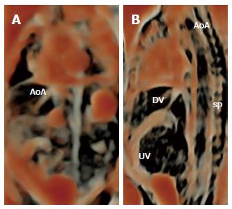
Figure 3 Normal fetal aortic arch at 28 wk of gestation.
4DUS echocardioangiography using HDliveTM application to the study of the great artery and veins: the AoA (A, image is rotated), the UV and the DV (B) are rendered with an enhanced quality resembling that of an angiographic study (sp, fetal spine). AoA: Aortic arch; UV: Umbilical vein; DV: Ductus venosus; sp: Fetal spine.
- Citation: Tonni G, Grisolia G, Santana EF, Júnior EA. Assessment of fetus during second trimester ultrasonography using HDlive software: What is its real application in the obstetrics clinical practice? World J Radiol 2016; 8(12): 922-927
- URL: https://www.wjgnet.com/1949-8470/full/v8/i12/922.htm
- DOI: https://dx.doi.org/10.4329/wjr.v8.i12.922


