Copyright
©The Author(s) 2016.
World J Radiol. Nov 28, 2016; 8(11): 895-901
Published online Nov 28, 2016. doi: 10.4329/wjr.v8.i11.895
Published online Nov 28, 2016. doi: 10.4329/wjr.v8.i11.895
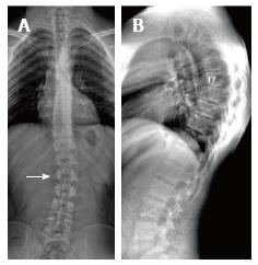
Figure 4 Twenty-three years old male patient with typical Scheuermann’s disease (Patient no: 7).
A: A scoliosis radiography demonstrating scoliosis with opening facing to right (white arrow) in lumbar axis; B. Lateral radiography of kyphosis with apex facing to T7 vertebra in thoracic spinal axis (Cobb angle 60.1°) and irregularities in thoracic endplates are shown.
- Citation: Gokce E, Beyhan M. Radiological imaging findings of scheuermann disease. World J Radiol 2016; 8(11): 895-901
- URL: https://www.wjgnet.com/1949-8470/full/v8/i11/895.htm
- DOI: https://dx.doi.org/10.4329/wjr.v8.i11.895


