Copyright
©The Author(s) 2016.
World J Radiol. Nov 28, 2016; 8(11): 868-879
Published online Nov 28, 2016. doi: 10.4329/wjr.v8.i11.868
Published online Nov 28, 2016. doi: 10.4329/wjr.v8.i11.868
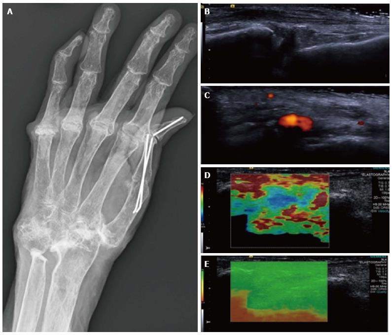
Figure 6 Synovitis at the carpus.
Radiograph (A) in a patient with long-standing rheumatoid arthritis and worsening wrist pain. The longitudinal B mode image (B) and colour Doppler image (C) show hypoechogenicity of the synovium with increased Doppler flow indicating an active synovitis. The corresponding longitudinal shear wave velocity elastogram (D) shows relatively uniformly soft synovium (blue colour) compared with the adjacent tissues which are more heterogeneous in their stiffness. The significance of this has not yet been adequately investigated. The elastogram is of good quality (E).
- Citation: Winn N, Lalam R, Cassar-Pullicino V. Sonoelastography in the musculoskeletal system: Current role and future directions. World J Radiol 2016; 8(11): 868-879
- URL: https://www.wjgnet.com/1949-8470/full/v8/i11/868.htm
- DOI: https://dx.doi.org/10.4329/wjr.v8.i11.868


