Copyright
©The Author(s) 2016.
World J Radiol. Nov 28, 2016; 8(11): 868-879
Published online Nov 28, 2016. doi: 10.4329/wjr.v8.i11.868
Published online Nov 28, 2016. doi: 10.4329/wjr.v8.i11.868
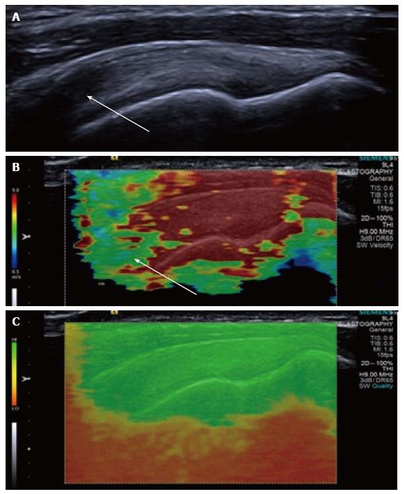
Figure 3 Longitudinal shear wave elastography of a normal supraspinatus tendon.
Normal B mode appearances (A) showing anisotropy (arrow) owing to the curved orientation of the tendon. The corresponding elastogram (B) shows heterogeneous stiffness at the region of anisotropy (arrow) and absence of measurements from deep in the humeral head. The quality map (C) shows a high quality elastogram in the tendon substance and poor quality in the bone, as would be expected for such a stiff structure with limited propagation of shear waves.
- Citation: Winn N, Lalam R, Cassar-Pullicino V. Sonoelastography in the musculoskeletal system: Current role and future directions. World J Radiol 2016; 8(11): 868-879
- URL: https://www.wjgnet.com/1949-8470/full/v8/i11/868.htm
- DOI: https://dx.doi.org/10.4329/wjr.v8.i11.868


