Copyright
©The Author(s) 2016.
World J Radiol. Nov 28, 2016; 8(11): 868-879
Published online Nov 28, 2016. doi: 10.4329/wjr.v8.i11.868
Published online Nov 28, 2016. doi: 10.4329/wjr.v8.i11.868
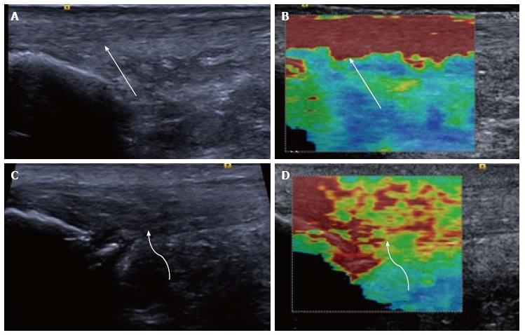
Figure 2 Longitudinal shear wave elastography of the Achilles tendon.
In a healthy volunteer the Achilles tendon is seen as smooth and homogeneous (arrow) on the B mode image (A) with a homogeneous elastogram (B, arrow). In a patient with symptomatic Achilles tendinopathy there is an alteration of the B mode echotexture with regions of hypoechogenicity (C, curved arrow) and dystrophic ossification at the calcaneal enthesis. The elastogram (D) is heterogeneous with regions of blue and yellow colouring (curved arrow) corresponding to a slower velocity and tendon softening.
- Citation: Winn N, Lalam R, Cassar-Pullicino V. Sonoelastography in the musculoskeletal system: Current role and future directions. World J Radiol 2016; 8(11): 868-879
- URL: https://www.wjgnet.com/1949-8470/full/v8/i11/868.htm
- DOI: https://dx.doi.org/10.4329/wjr.v8.i11.868


