Copyright
©The Author(s) 2016.
World J Radiol. Nov 28, 2016; 8(11): 857-867
Published online Nov 28, 2016. doi: 10.4329/wjr.v8.i11.857
Published online Nov 28, 2016. doi: 10.4329/wjr.v8.i11.857
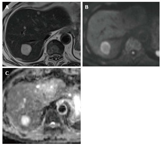
Figure 1 A 65-year-old man with metastatic tumor in the liver from colorectal carcinoma.
A: T2-weighted imaging shows an obvious hyperintense lesion on segment VII (arrow); B: DWI (b-value of 800 s/mm2) shows hyperintensity; C: Apparent diffusion coefficient map also shows hyperintensity. This finding mimics that for hemangioma. DWI: Diffusion-weighted imaging.
- Citation: Saito K, Tajima Y, Harada TL. Diffusion-weighted imaging of the liver: Current applications. World J Radiol 2016; 8(11): 857-867
- URL: https://www.wjgnet.com/1949-8470/full/v8/i11/857.htm
- DOI: https://dx.doi.org/10.4329/wjr.v8.i11.857


