Copyright
©The Author(s) 2016.
World J Radiol. Nov 28, 2016; 8(11): 851-856
Published online Nov 28, 2016. doi: 10.4329/wjr.v8.i11.851
Published online Nov 28, 2016. doi: 10.4329/wjr.v8.i11.851
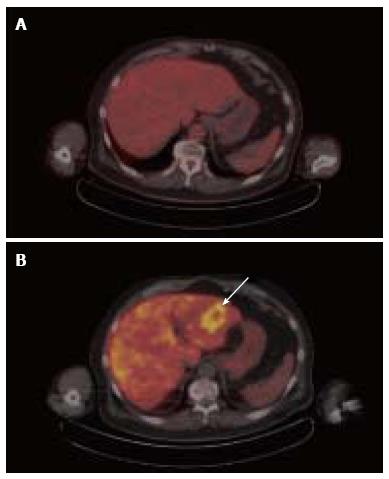
Figure 3 Corresponding 18F-fluorodeoxy-D-glucose positron emission tomography/computed tomography (A) and 18F-fluorocholine positron emission tomography/computed tomography (B) images of hepatocellular carcinoma obtained from the same patient on different days.
The tumor is not at all evident on transaxial images of the liver from FDG PET/CT (A). Corresponding transaxial images of the liver from 18F-fluorocholine PET/CT (B) shows a 5-cm diameter circumscribed area of increased uptake in the left hepatic lobe (B) This tumor contained within the left hepatic lobe was histopathologically confirmed to be a well-differentiated HCC. FDG: 18F-fluorodeoxy-D-glucose; PET: Positron emission tomography; CT: Computed tomography; HCC: Hepatocellular carcinoma.
- Citation: Kwee SA, Lim J. Metabolic positron emission tomography imaging of cancer: Pairing lipid metabolism with glycolysis. World J Radiol 2016; 8(11): 851-856
- URL: https://www.wjgnet.com/1949-8470/full/v8/i11/851.htm
- DOI: https://dx.doi.org/10.4329/wjr.v8.i11.851


