Copyright
©The Author(s) 2016.
World J Radiol. Oct 28, 2016; 8(10): 819-828
Published online Oct 28, 2016. doi: 10.4329/wjr.v8.i10.819
Published online Oct 28, 2016. doi: 10.4329/wjr.v8.i10.819
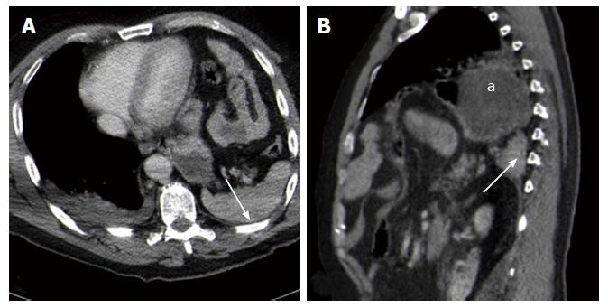
Figure 9 Computed tomography findings in blunt diaphragmatic lesions - dependent viscera sign.
These 3 mm thick multiplanar axial (A) and coronal (B) reconstructions show the spleen (arrows) and the stomach (a) lying on the posterior chest wall, without lung parenchyma interposition. This alteration represents the so-called “dependent viscera sign”.
- Citation: Bonatti M, Lombardo F, Vezzali N, Zamboni GA, Bonatti G. Blunt diaphragmatic lesions: Imaging findings and pitfalls. World J Radiol 2016; 8(10): 819-828
- URL: https://www.wjgnet.com/1949-8470/full/v8/i10/819.htm
- DOI: https://dx.doi.org/10.4329/wjr.v8.i10.819


