Copyright
©The Author(s) 2016.
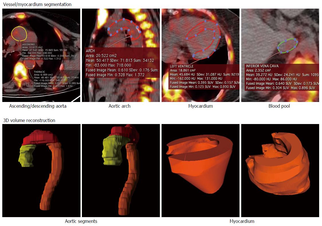
Figure 1 Volume of interest placement and 3-dimensional rendering of the vascular segments and of the myocardium.
Volumes of interest were constructed by placing sequential region of interest on the transaxial CT slices, carefully adjusting the edges so as to include the entire vessel/myocardium (top panels). The figure depicts the 3D rendering of the three aortic segments and of the heart (bottom panels). 3D: 3-dimensional; CT: Computed tomography.
- Citation: Fiz F, Morbelli S, Bauckneht M, Piccardo A, Ferrarazzo G, Nieri A, Artom N, Cabria M, Marini C, Canepa M, Sambuceti G. Correlation between thoracic aorta 18F-natrium fluoride uptake and cardiovascular risk. World J Radiol 2016; 8(1): 82-89
- URL: https://www.wjgnet.com/1949-8470/full/v8/i1/82.htm
- DOI: https://dx.doi.org/10.4329/wjr.v8.i1.82


