Copyright
©The Author(s) 2016.
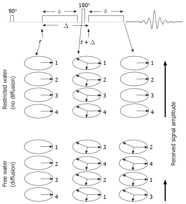
Figure 1 Schematic representation of the pulsed field gradient pulse sequence.
In this description we assume that we start the sequence with a sample containing only four in-phase spins labelled with 1, 2, 3 and 4. In the absence of diffusion, the first gradient pulse causes dephasing of the spins. The 180° radio-frequency pulse reverses the sign of the phase angle and thus after the second gradient pulse all spins are in phase which gives a maximum echo signal. In the presence of diffusion, spins go through a random walk process resulting in a distribution of phases. This in turn results in poorer refocusing of the spins and thus, a smaller echo signal.
- Citation: Jafar MM, Parsai A, Miquel ME. Diffusion-weighted magnetic resonance imaging in cancer: Reported apparent diffusion coefficients, in-vitro and in-vivo reproducibility. World J Radiol 2016; 8(1): 21-49
- URL: https://www.wjgnet.com/1949-8470/full/v8/i1/21.htm
- DOI: https://dx.doi.org/10.4329/wjr.v8.i1.21


