Copyright
©The Author(s) 2016.
World J Radiol. Jan 28, 2016; 8(1): 109-116
Published online Jan 28, 2016. doi: 10.4329/wjr.v8.i1.109
Published online Jan 28, 2016. doi: 10.4329/wjr.v8.i1.109
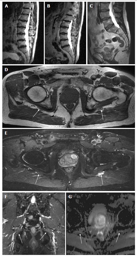
Figure 3 Left sciatic chronic neuropathy.
A 65-year-old man with prior left pelvic bone fracture and persistent left radicular pain. EMG was negative. MR imaging of LS spine obtained 12 mo (A) and 6 mo (B) before the MRN LS plexus examination (C-F) shows multilevel mild degenerative changes. Axial T1W (D), corresponding T2 SPAIR (E), 3D IR TSE (F) and axial DTI tensor (G) images show asymmetric mild increased signal of the left sciatic nerve with mild fatty infiltration (arrows). MR: Magnetic resonance; LS: Lumbosacral; T: Tesla; MRN: Magnetic resonance neurography; IR: Inversion recovery; 3D: 3-Dimensional; DTI: Diffusion tensor imaging; SPAIR: Spectral adiabatic inversion recovery; EMG: Electromyogram.
- Citation: Chhabra A, Farahani SJ, Thawait GK, Wadhwa V, Belzberg AJ, Carrino JA. Incremental value of magnetic resonance neurography of Lumbosacral plexus over non-contributory lumbar spine magnetic resonance imaging in radiculopathy: A prospective study. World J Radiol 2016; 8(1): 109-116
- URL: https://www.wjgnet.com/1949-8470/full/v8/i1/109.htm
- DOI: https://dx.doi.org/10.4329/wjr.v8.i1.109


