Copyright
©The Author(s) 2016.
World J Radiol. Jan 28, 2016; 8(1): 109-116
Published online Jan 28, 2016. doi: 10.4329/wjr.v8.i1.109
Published online Jan 28, 2016. doi: 10.4329/wjr.v8.i1.109
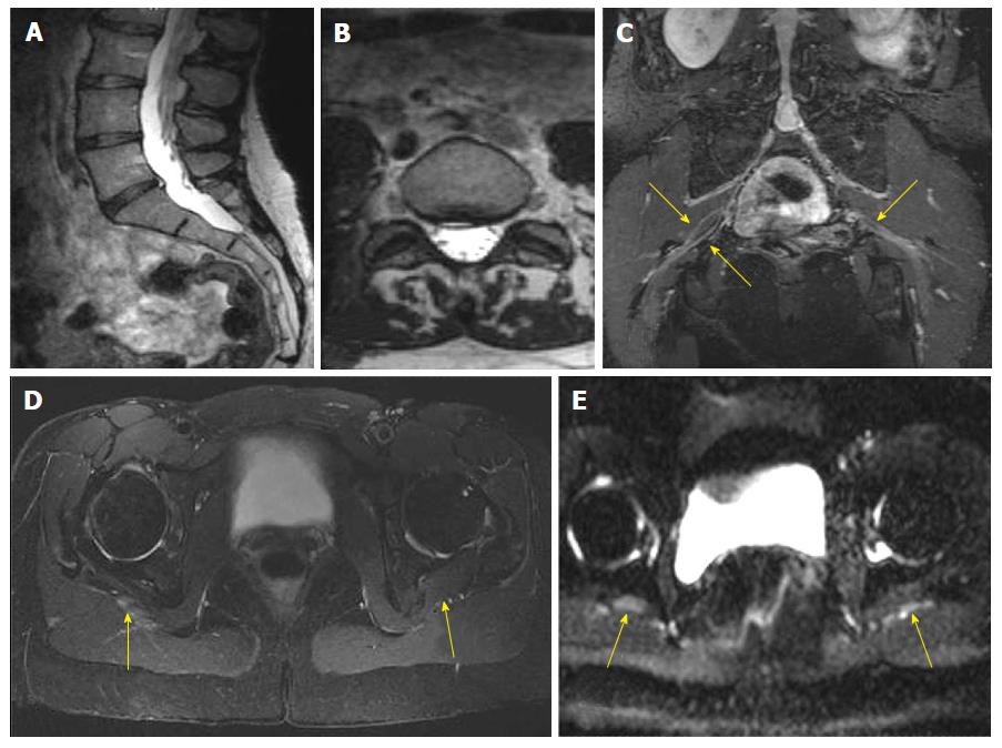
Figure 2 Right piriformis syndrome.
A 42-year-old woman with 2 yr history of intermittent partial foot drops and right gluteal pain. EMG was negative. Outside MR imaging of LS spine was reported normal, except small disc herniation at L5-S1 level. MRN LS plexus (A-E) shows small annular fissure and right paracentral disc herniation at L5-S1 level (A, B) on 3D TSE imaging of lumbar spine. Coronal 3D IR TSE (C) shows bilateral split sciatic nerves (right > left, arrows). Axial T2 SPAIR (D) image shows mildly enlarged and hyperintense right sciatic nerve (arrows), immediately outside the greater sciatic notch. Axial DTI (E) confirms more conspicuity of the right sciatic nerve abnormality (arrows). MR: Magnetic resonance; LS: Lumbosacral; MRN: Magnetic resonance neurography; IR: Inversion recovery; 3D: 3-Dimensional; DTI: Diffusion tensor imaging; SPAIR: Spectral adiabatic inversion recovery; EMG: Electromyogram; TSE: Turbo spin echo..
- Citation: Chhabra A, Farahani SJ, Thawait GK, Wadhwa V, Belzberg AJ, Carrino JA. Incremental value of magnetic resonance neurography of Lumbosacral plexus over non-contributory lumbar spine magnetic resonance imaging in radiculopathy: A prospective study. World J Radiol 2016; 8(1): 109-116
- URL: https://www.wjgnet.com/1949-8470/full/v8/i1/109.htm
- DOI: https://dx.doi.org/10.4329/wjr.v8.i1.109


