Copyright
©The Author(s) 2016.
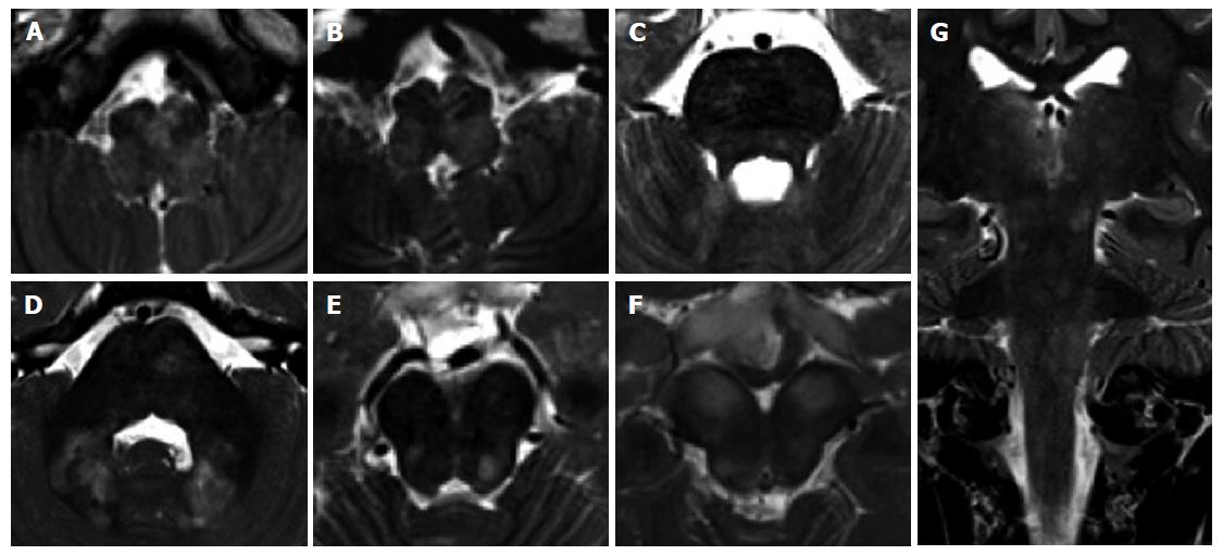
Figure 18 Unidentified bright objects in neurofibromatosis type 1.
A 11-year-old girl with NF1. Multiple bilateral symmetric or asymmetric T2 hyperintense lesions, diffuse and confluent across the brainstem, in the cerebellar white matter and at supratentorial level may be encountered in these patients. These lesions do not enhance after contrast agent injection. Axial (A-F) and coronal (G) T2 weighted images of the medulla (A,B), pons (C,D) and midbrain (E,F) are presented. Coronal image (G) shows the involvement of infra- and supra-tentorial areas.
- Citation: Quattrocchi CC, Errante Y, Rossi Espagnet MC, Galassi S, Della Sala SW, Bernardi B, Fariello G, Longo D. Magnetic resonance imaging differential diagnosis of brainstem lesions in children. World J Radiol 2016; 8(1): 1-20
- URL: https://www.wjgnet.com/1949-8470/full/v8/i1/1.htm
- DOI: https://dx.doi.org/10.4329/wjr.v8.i1.1


