Copyright
©The Author(s) 2015.
World J Radiol. Sep 28, 2015; 7(9): 294-305
Published online Sep 28, 2015. doi: 10.4329/wjr.v7.i9.294
Published online Sep 28, 2015. doi: 10.4329/wjr.v7.i9.294
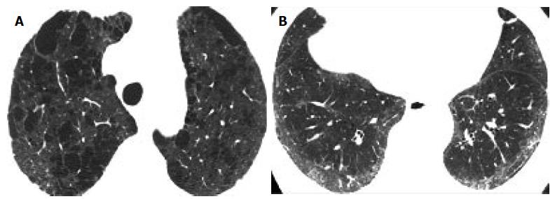
Figure 2 Centrilobular emphysema and nonspecific interstitial pneumonia-pattern of pulmonary fibrosis.
A: High resolution computed tomography (HRCT) at the level of the upper lobes shows extensive predominantly centrilobular emphysema; B: HRCT of the same patient as in A, at the level of the lower lobes shows subpleural fine fibrosis characterized mainly by ground glass opacity, reticular pattern non-otherwise specified and absence of honeycombing. The HRCT pattern is consistent more with nonspecific interstitial pneumonia.
- Citation: Oikonomou A, Mintzopoulou P, Tzouvelekis A, Zezos P, Zacharis G, Koutsopoulos A, Bouros D, Prassopoulos P. Pulmonary fibrosis and emphysema: Is the emphysema type associated with the pattern of fibrosis? World J Radiol 2015; 7(9): 294-305
- URL: https://www.wjgnet.com/1949-8470/full/v7/i9/294.htm
- DOI: https://dx.doi.org/10.4329/wjr.v7.i9.294


