Copyright
©The Author(s) 2015.
World J Radiol. Sep 28, 2015; 7(9): 253-265
Published online Sep 28, 2015. doi: 10.4329/wjr.v7.i9.253
Published online Sep 28, 2015. doi: 10.4329/wjr.v7.i9.253
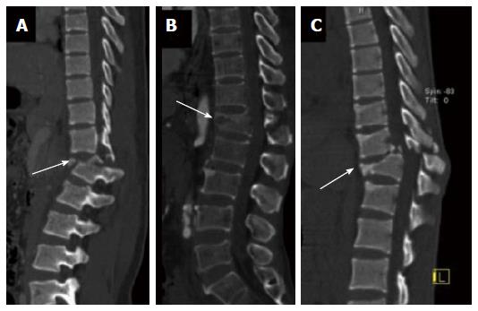
Figure 9 Type B2 flexion distraction injury.
Sagittal computed tomography (CT) image (A), sagittal and coronal CT images (B) and sagittal and axial CT images (C) show transverse bicolumn fracture (B 2.1) [arrow in (A)], flexion-spondylolysis (B 2.2) [arrow in (B)] and (C) flexion- distraction with type A fracture (B 2.3) (arrow).
- Citation: Gamanagatti S, Rathinam D, Rangarajan K, Kumar A, Farooque K, Sharma V. Imaging evaluation of traumatic thoracolumbar spine injuries: Radiological review. World J Radiol 2015; 7(9): 253-265
- URL: https://www.wjgnet.com/1949-8470/full/v7/i9/253.htm
- DOI: https://dx.doi.org/10.4329/wjr.v7.i9.253


