Copyright
©The Author(s) 2015.
World J Radiol. Sep 28, 2015; 7(9): 253-265
Published online Sep 28, 2015. doi: 10.4329/wjr.v7.i9.253
Published online Sep 28, 2015. doi: 10.4329/wjr.v7.i9.253
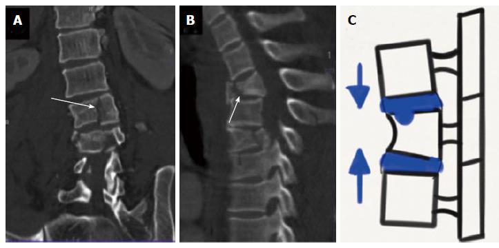
Figure 6 Type A2 compression injuries.
Coronal computed tomography (CT) image (A), sagittal CT image (B) and schematic diagram (C) show (A) sagittal split (A2.1), (B) coronal split (A 2.2) and (C) pincer type injury (A 2.3).
- Citation: Gamanagatti S, Rathinam D, Rangarajan K, Kumar A, Farooque K, Sharma V. Imaging evaluation of traumatic thoracolumbar spine injuries: Radiological review. World J Radiol 2015; 7(9): 253-265
- URL: https://www.wjgnet.com/1949-8470/full/v7/i9/253.htm
- DOI: https://dx.doi.org/10.4329/wjr.v7.i9.253


