Copyright
©The Author(s) 2015.
World J Radiol. Jul 28, 2015; 7(7): 180-183
Published online Jul 28, 2015. doi: 10.4329/wjr.v7.i7.180
Published online Jul 28, 2015. doi: 10.4329/wjr.v7.i7.180
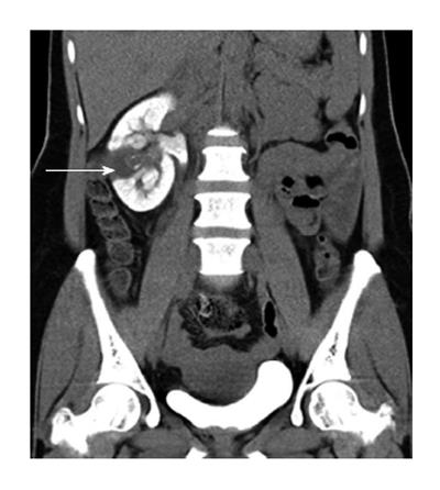
Figure 3 Delayed oblique coronal multiplanar reformat MDCT image shows the lesion as a filling defect within the collecting system.
The lesion and laterally placed cortical cyst are inseparable. Additionally, persistent nephrogram is noted.
- Citation: Bhaya A, Shinde AP. Isolated renal hydatid presenting as a complex renal lesion followed by spontaneous hydatiduria. World J Radiol 2015; 7(7): 180-183
- URL: https://www.wjgnet.com/1949-8470/full/v7/i7/180.htm
- DOI: https://dx.doi.org/10.4329/wjr.v7.i7.180


