Copyright
©The Author(s) 2015.
World J Radiol. Jul 28, 2015; 7(7): 180-183
Published online Jul 28, 2015. doi: 10.4329/wjr.v7.i7.180
Published online Jul 28, 2015. doi: 10.4329/wjr.v7.i7.180
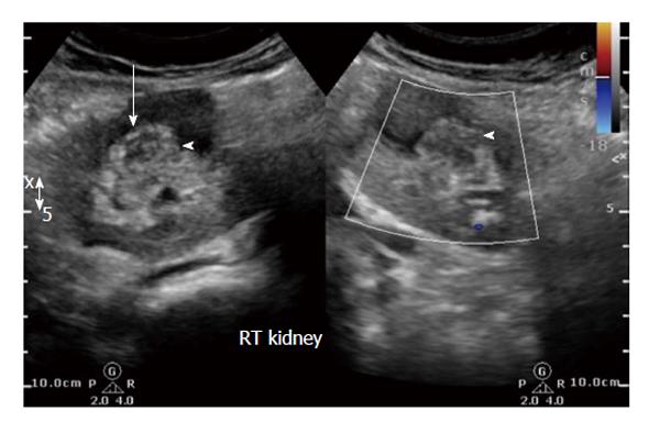
Figure 1 Initial ultrasound image reveals heteroechoic indeterminate renal sinus/renal pelvic lesion (white arrow).
Doppler revealed negligible vascularity.
- Citation: Bhaya A, Shinde AP. Isolated renal hydatid presenting as a complex renal lesion followed by spontaneous hydatiduria. World J Radiol 2015; 7(7): 180-183
- URL: https://www.wjgnet.com/1949-8470/full/v7/i7/180.htm
- DOI: https://dx.doi.org/10.4329/wjr.v7.i7.180


