Copyright
©The Author(s) 2015.
World J Radiol. Jul 28, 2015; 7(7): 149-156
Published online Jul 28, 2015. doi: 10.4329/wjr.v7.i7.149
Published online Jul 28, 2015. doi: 10.4329/wjr.v7.i7.149
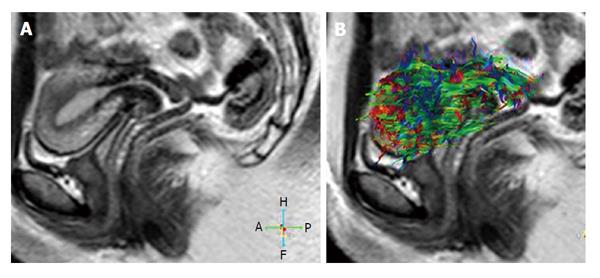
Figure 4 Thirty two years old volunteer.
A: Sagittal T2-weighted image of a normal uterus; B: 3D whole tractography image of the normal uterus. Red colors represent a right-left orientation, blue represents a cranio-caudal orientation and green represents an antero-posterior orientation of diffusion. Changes in the intensity of the color represent different strengths of anisotropy.
- Citation: Kara Bozkurt D, Bozkurt M, Nazli MA, Mutlu IN, Kilickesmez O. Diffusion-weighted and diffusion-tensor imaging of normal and diseased uterus. World J Radiol 2015; 7(7): 149-156
- URL: https://www.wjgnet.com/1949-8470/full/v7/i7/149.htm
- DOI: https://dx.doi.org/10.4329/wjr.v7.i7.149


