Copyright
©The Author(s) 2015.
World J Radiol. Jul 28, 2015; 7(7): 149-156
Published online Jul 28, 2015. doi: 10.4329/wjr.v7.i7.149
Published online Jul 28, 2015. doi: 10.4329/wjr.v7.i7.149
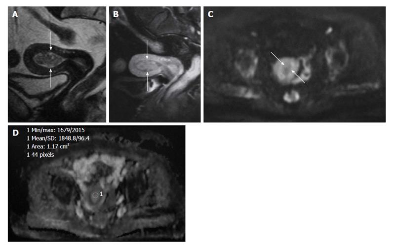
Figure 3 A 42-year-old woman with endometrial polyp.
A: Hypointense polyp in the endometrial cavity on sagittal T2-weighted image mimicking low grade endometrial carcinoma (arrows); B: Sagittal contrast-enhanced T1-weighted image with fat suppression shows enhancing endometrial polyp (arrows); C: On the axial DWI (b = 1000 s/mm2) image, the mass is hypointense clearly excluding malignancy (arrows); D: Corresponding axial apparent diffusion coefficient (ADC) map. The ADC value within the mass is 1.85 × 10⁻³ mm2/s.
- Citation: Kara Bozkurt D, Bozkurt M, Nazli MA, Mutlu IN, Kilickesmez O. Diffusion-weighted and diffusion-tensor imaging of normal and diseased uterus. World J Radiol 2015; 7(7): 149-156
- URL: https://www.wjgnet.com/1949-8470/full/v7/i7/149.htm
- DOI: https://dx.doi.org/10.4329/wjr.v7.i7.149


